Note: We do not currently have a health liaison for this disorder. If you would like to volunteer, please contact president@samoyedhealthfoundation.org and we will be happy to answer any of your questions. For a description of the position, please click on disorder health information liaison or health information reviewer.
Thank you to Joy Ritter for putting together this article for SCARF.
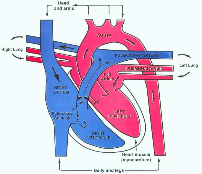
In normal circulation, the ventricle on the right side of the heart pumps blood to the lungs where it picks up oxygen. The oxygenated blood then returns to the left side of the heart and is pumped to the rest of the body.
As the blood leaves the right ventricle it flows through the pulmonic valve into the pulmonary artery which goes to the lungs. With pulmonic stenosis (PS), there is a narrowing (stenosis) or obstruction in this path. This is usually due to a malformation of the pulmonic valve ("pulmonic valve dysplasia"), but the abnormality may be just above or below the valve as well.
The heart must work harder to pump the blood past the obstruction. This extra workload causes the right ventricle to thicken. The extent to which a dog may be affected depends on the degree of narrowing of the valve area. With severe stenosis the increased workload on the heart may lead to right-side congestive heart failure.
See a diagram of the heart showing the pulmonic stenosis.
Signs and Symptoms
- Heart murmur, lower left quadrant of heart
- In symptomatic cases, exercise intolerance, lethargy, lack of appetite
- Pale gums, fainting spells
- In severe cases, right side congestive heart failure
Causes
- Congenital (present at birth due to developmental problems or genetics). Inheritable in some breeds as a polygenic threshold trait (complex inheritance controlled by a number of genes). It appears to be inheritable in Samoyeds, although this has yet to be confirmed in formal scientific studies. It is one of the most common heart defects seen in Samoyeds.
Risk Factors
Parent(s) and/or sibling(s) with disease.
Diagnostic Tests
- Manual Auscultation (listening with a stethoscope) by board certified cardiologist, followed by
- Color Doppler echocardiography (a type of ultrasound) to definitively diagnose the disease
- Chest Xrays
- Once a diagnosis has been made, a cardiac catherization can be done to measure the pressure gradient between the right ventricle and the pulmonary artery, to determine the severity of the obstruction.
Treatment Guidelines
Note: Treatment of animals should only be performed by a licensed veterinarian. Veterinarians should consult the current literature and current pharmacological formularies before initiating any treatment protocol.
- Mild forms, no treatment necessary.
- Moderate to severe forms, relief of symptoms as necessary (diuretics, blood pressure medications for heart failure relief and slow heart function), under the direction of a veterinary cardiologist, if possible.
- Surgery (for example, balloon valvuloplasty) is possible to repair the valve in some cases.
- Prognosis for mild forms, good. However, should NOT be bred, as it is a congenital condition.
Management
From an owner's perspective:
The mild form of PS is self limiting. Dogs should be spayed/neutered to prevent accidental pregnancy. Nutritional advice would be to keep the dog at optimum weight. Overweight dogs put a strain on the heart (already stressed). Underweight due to nutritional challenges also puts stress on the heart. Treat symptoms as they arise and have periodic (annual) checkups with a cardio vet to access progression of the disease. Allow the dog to self limit his exercise. No FORCED exercise (pulling, sledding, jogging, etc.).
References
- Understanding Canine Pulmonic
Stenosis
from the Matthew J. Ryan Veterinary Hospital, University of
Pennsylvania School of Veterinary Medicine.

- Pulmonic
Stenosis
from the Canine Inherited Disorders Database

Suggested Links
- http://www.vet.upenn.edu/veterinary-hospitals/ryan-veterinary-hospital/services/cardiology
Search for 'pulmonic stenosis'. This site has a wealth of
information on it, as well as slide presentations.
- https://www.ofa.org/diseases/other-diseases/cardiac-disease -- This link has good
descriptions of the cardiac physical and ultrasound exams.
- Pulmonic
Stenosis
from the Canine Inherited Disorders Database
- http://www.mirage-samoyeds.com/heart.htm --
Samoyed State of Heart by Cheri Hollenback

